Checklist for Understanding Acemannan Mechanisms
Links acemannan's acetylated, mannose-rich structure to immune signaling, wound and bone repair, and gut fermentation.

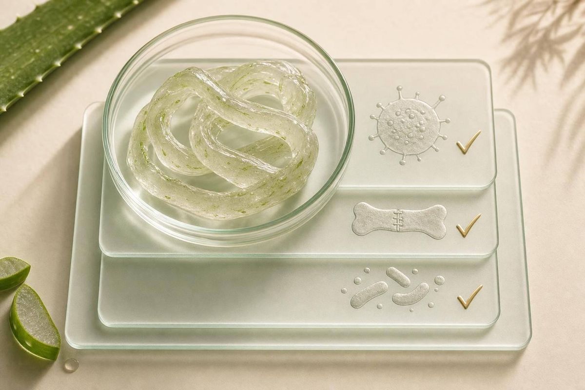
Here’s the short answer: acemannan works because of its structure - mainly its acetyl groups, mannose-rich β(1→4) backbone, and molecular size. Those features shape how it interacts with immune cells, repair cells, bone-forming cells, and gut microbes.
If I had to boil this acemannan research overview down fast, I’d keep these points:
- Structure comes first. Acemannan is mostly mannose (~93%), with small amounts of glucose (~3%) and galactose (~3%).
- Acetylation matters a lot. When acemannan is fully deacetylated, it does not trigger cell proliferation or VEGF and Collagen I expression in the same way.
- Size matters too. Native acemannan can reach ~2,000 kDa, while some processed forms are closer to ~80 kDa, which can shift activity.
- Immune effects center on macrophages and dendritic cells. Reported markers include IL-1, IL-6, TNF-α, NO, phagocytosis, and M2 polarization.
- Repair effects center on fibroblasts, keratinocytes, and bone cells. Main pathways include AKT/mTOR, EGFR/PKC, and osteogenic markers like ALP, BMP-2, and BMP-4.
- Gut effects depend on fermentation. Acemannan reaches the colon, where microbes turn it into SCFAs such as acetate, propionate, and butyrate.
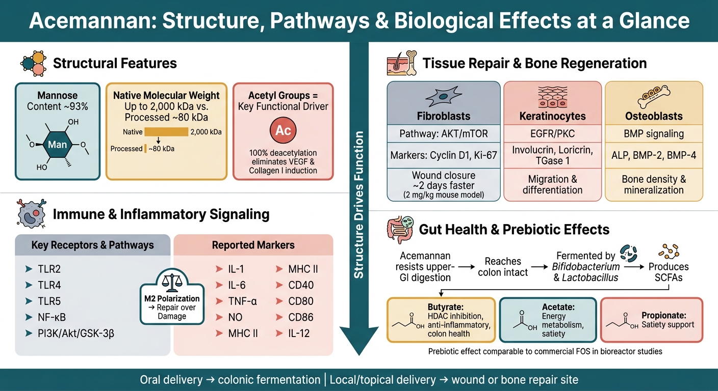
Acemannan: Structure, Pathways & Biological Effects at a Glance
Quick Comparison
| Area | What to check | Main point |
|---|---|---|
| Structure | Acetylation, β(1→4) polymannan backbone, MW | Structure shapes activity |
| Immune signaling | TLRs, NF-κB, cytokines, M2 shift | Effects vary by cell type |
| Tissue repair | Fibroblasts, collagen, VEGF, AKT/mTOR | Supports wound-healing signals |
| Bone | ALP, BMP-2, BMP-4, mineralization | Linked to osteogenic activity |
| Gut | SCFAs, microbiome shifts, barrier repair | Works partly through colonic fermentation |
Bottom line: this article is a step-by-step way to connect chemistry → signaling pathways → biological effects, while keeping cell studies, animal data, and human data separate.
sbb-itb-b88dd21
Checklist 1: Confirm the Structural Features That Drive Activity
Check the Core Chemistry: Acetylated β(1→4)-Linked Polymannan
Start here: confirm acemannan's backbone, branches, and acetyl groups. Those parts decide how it acts.
Acemannan is a water-soluble polysaccharide with a β(1→4)-linked mannose backbone, minor glucose content, and α(1→6) galactose branches. Its main sugars are about 93% mannose, 3% glucose, and 3% galactose.
The acetyl groups matter a lot. They set acemannan apart from other aloe polysaccharides because they are the only non-sugar functional groups in the molecule. They also drive binding with immune receptors, including mannose receptors, TLR4, and TLR5 on macrophages and dendritic cells.
To confirm that identity, researchers use 1H-NMR and IR spectra to detect acetyl groups and verify sugar ratios.
Once that structure is confirmed, the next job is to check how acetylation and molecular size affect bioactivity.
Check Why Acetylation and Molecular Size Matter
Acetylation and molecular size are the main structural variables behind receptor interaction, solubility, and downstream bioactivity.
Native acemannan in fresh Aloe vera gel can reach molecular weights of up to 2,000 kDa (2 million Daltons). That higher molecular weight is linked to stronger immune stimulation. Processed versions, such as Modified Aloe Polysaccharide (MAP), average around 80 kDa, which changes which effects stand out most.
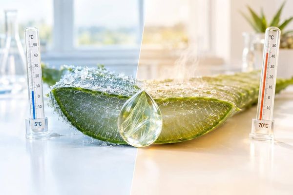
Acetylation also changes how the molecule behaves in a direct way. Research shows that 100% deacetylated acemannan fails to induce cell proliferation or the expression of VEGF and Collagen I, while acetylated acemannan does. Deacetylation lowers solubility and shifts the molecule into a more ordered structure with less receptor activity. Processing can weaken activity too. For example, spray-drying can lower acetylation by up to 70%.
| Structural Feature | Effect on Acemannan |
|---|---|
| More acetylation | Maintains water solubility, 3D structure, and immune/tissue repair activity |
| Deacetylation | Reduces bioactivity, lowers solubility, and creates a more rigid structure |
| High molecular weight (~2,000 kDa) | Supports stronger immune activity |
| Lower-MW fragments | Can increase specific effects such as vaccine efficacy and macrophage activation |
These features help explain why acemannan shows some of its strongest effects in immune, repair, and gut pathways.
Checklist 2: Track the Best-Supported Immune and Inflammatory Mechanisms
Check Macrophage Activation, Cytokines, and Toll-Like Receptor Signaling
Once acemannan's structure is confirmed, the next step is to track the immune effects with the strongest support. In plain English: after structure, look at the downstream immune signals that show up most often in the research.
Start with macrophages. Acemannan can trigger release of IL-1, IL-6, TNF-α, and NO, with stronger effects in the presence of IFN-γ. It also supports phagocytosis and Candida-killing activity.
It doesn’t stop at activation alone. Acemannan also promotes M2 polarization and suppresses LPS-induced M1 polarization, which points signaling more toward repair than damage.
Then trace the receptor-level signals behind those effects. Check TLR5 in human gingival fibroblasts and the downstream PI3K/Akt/GSK-3β pathway. It also makes sense to check dendritic-cell maturation markers, including MHC II, CD40, CD54, CD80, CD86, and IL-12.
Check NF-κB Regulation, T-Cell Support, and Antiviral Relevance
NF-κB does not behave the same way in every cell type. That detail matters here.
In gingival fibroblasts, acemannan activates TLR5/NF-κB and increases IL-6 and IL-8. In keratinocytes, it does almost the opposite and suppresses TNF-α-induced NF-κB signaling.
From there, check whether those signals feed into adaptive immune activity. T-cell support appears to be indirect through dendritic-cell maturation. Some studies also report dose-dependent T-lymphocyte proliferation and cytotoxicity.
For antiviral evidence, the cleanest way to frame the mechanism is host-defense support. Acemannan activates macrophages and T-cells rather than directly targeting the pathogen.
| Mechanism | Key Markers to Check | Pathway |
|---|---|---|
| Macrophage activation | IL-1, IL-6, TNF-α, NO, M2 polarization | PI3K/Akt/GSK-3β |
| Dendritic cell maturation | MHC II, CD40, CD54, CD80, CD86, IL-12 | Mannose receptor agonism |
| NF-κB regulation | IL-6, IL-8 (activation) or reduced TNF-α-induced NF-κB signaling | TLR5 / TNF-α inhibition |
| T-cell support | Cytotoxic T-lymphocyte proliferation | Interferon-related activity |
Prioritize studies using native, acetylated acemannan.
Checklist 3: Connect Acemannan to Tissue Repair, Wound Healing, and Bone Regeneration
Check Fibroblast Activity, AKT/mTOR Signaling, and Collagen Support
Acemannan helps skin wounds heal by driving protein translation through the AKT/mTOR pathway. In plain terms, it nudges repair cells to grow, move, and get to work. It stimulates fibroblast proliferation and migration, and in gingival fibroblasts it triggers production of Type I collagen, KGF-1, and VEGF at 2–16 mg/mL.
The main pathway here is AKT/mTOR. Acemannan promotes translation of cyclin D1, which pushes cells through the cell cycle. When researchers used rapamycin, the effect was blocked, which confirmed the pathway’s role. In a mouse skin wound model, acemannan at 2 mg/kg sped up wound closure by about 2 days versus controls.
That said, fibroblasts are only part of the repair story. Epithelial closure depends on keratinocytes, and they rely on EGFR/PKC signaling to support proliferation, migration, and differentiation.
| Pathway | Primary Cell Type | Key Markers | Repair Outcome |
|---|---|---|---|
| AKT/mTOR | Fibroblasts | Cyclin D1, Ki-67 | Proliferation, wound closure |
| EGFR/PKC | Keratinocytes | Involucrin, Loricrin, TGase 1 | Migration, differentiation |
| Growth factor release | Gingival fibroblasts | KGF-1, VEGF, Type I collagen | ECM deposition, re-epithelialization |
Check Osteoblast and Bone Remodeling Mechanisms
The repair effect doesn’t stop at soft tissue. It also reaches mineralized tissue, where similar signals act in osteoblast-like cells. Acemannan supports osteogenic repair through bone marrow stromal cells and dental pulp cells. It stimulates their proliferation, differentiation, and mineralization, and it upregulates VEGF and its receptors during osteoblast differentiation to support vascular supply at the repair site.
Animal and clinical models also show more bone formation, higher bone density, and better healing after acemannan treatment.
When reviewing bone remodeling activity, look for markers such as:
- Alkaline phosphatase
- BMP-2
- BMP-4
- Bone volume
- Bone density
Checklist 4: Understand Gut and Microbiome-Related Mechanisms
Check Prebiotic Fermentation and Short-Chain Fatty Acid Production
After repair and bone, the next place to look is the gut. That’s where acemannan works as both a fermentable substrate and an immune modulator.
Because acemannan resists upper-GI digestion, it reaches the colon mostly intact. Once it gets there, it becomes food for gut bacteria, especially Bifidobacterium and Lactobacillus species. That selective support is why acemannan is treated as a prebiotic.
When gut microbes ferment acemannan, they produce short-chain fatty acids, or SCFAs, mainly acetate, propionate, and butyrate. In bioreactor studies built to mimic human colon conditions, acemannan increased acetate concentrations by a clear margin. In that same model, its effect on bacterial growth was on par with commercial fructooligosaccharides (FOS).
Butyrate matters a lot here. It acts as a histone deacetylase (HDAC) inhibitor, which helps regulate inflammatory gene expression and supports colon health. So while all three SCFAs play a role, butyrate tends to get the most attention.
In plain terms, acemannan’s prebiotic effect depends on having enough Bifidobacterium and Lactobacillus in the gut to convert it into SCFAs such as butyrate.
| SCFA | Primary Proposed Benefit |
|---|---|
| Butyrate | Colon health, anti-inflammatory signaling, HDAC inhibition |
| Acetate | Energy metabolism and satiety regulation |
| Propionate | Satiety support |
Fermentation is only one side of the picture. Acemannan also appears to shape immune activity inside the gut.
Check Gut Immune Signaling and Local Inflammation
Beyond fermentation, acemannan also affects local gut immune signaling.
It interacts with gut immune cells, including macrophages and dendritic cells. More specifically, it binds to Toll-like receptor 4 (TLR-4) and TLR-2, which triggers innate immune signaling tied to control of local inflammation.
Acemannan also seems to support intestinal barrier repair. In a 2021 mouse study of an Aloe gel-based formula, oral use normalized both pro-inflammatory cytokines (TNF-α, IL-6) and anti-inflammatory cytokines (IL-4, IL-10) in adipose tissue. That shift correlated with higher relative abundance of gut bacteria such as Bacteroides, Butyricimonas, Ruminococcus, and Mucispirillum.
M2 polarization is part of this repair response. It supports intestinal barrier repair and shifts the local setting away from inflammation and toward repair. Oral consumption is also linked to re-epithelialization at injured sites in the small intestine and colon.
That said, most of the evidence here comes from in vitro bioreactor work and animal studies.
Conclusion: Use the Checklist to Link Structure, Pathways, and Outcomes
These four checklists tie acemannan's structure to its immune, repair, and gut health benefits. The same pattern shows up in every section.
Key Takeaways to Review Before You Finish
The main point is simple: structure drives function. Acemannan's acetylated β(1,4)-linked polymannan backbone sits at the center of its activity. When processing strips away acetyl groups, bioactivity drops.
From there, the pathways split based on where acemannan is used and how it's delivered. The table below maps each mechanism to its pathway and the part of the structure doing the heavy lifting:
| Mechanism Category | Key Pathways | Primary Structural Driver |
|---|---|---|
| Immune Modulation | TLR2, TLR5, NF-κB, PI3K/Akt/GSK-3β | Acetylation and mannose-rich backbone |
| Tissue & Bone Repair | AKT/mTOR, PI3K/Akt | Porosity and acetylation |
| Gut Health | SCFA production, satiety signaling | β(1,4)-linked polymannan |
Oral acemannan works through colonic fermentation. Local biomaterials work at the wound or defect site. That difference in delivery is why the checklist separates oral and local use.
For a deeper mechanism review, see Advanced Acemannan.
FAQs
Why does acetylation matter so much?
Acetylation is what gives acemannan much of its punch. Its acetyl groups are the only non-sugar functional parts of the molecule, and they play a big role in shaping its biological activity, structure, and physicochemical properties.
Take those acetyl groups away, and acemannan changes in a big way. When it becomes deacetylated, it loses much of its therapeutic potential, including its ability to support cell maturation, immune activation, and the production of VEGF and Type I Collagen needed for tissue repair.
Does molecular weight change acemannan’s effects?
Yes. Molecular weight has a big effect on acemannan’s biological activity.
Native acemannan ranges from 30 kDa to 2 million Daltons, but its immunomodulating effects seem strongest in medium-sized chains, usually around 5 to 400 kDa.
Some studies also suggest that smaller fragments under 100 kDa may do more to support cell proliferation and tissue regeneration.
Put simply, chain size helps shape how the body processes and uses acemannan.
Is oral acemannan different from local use?
Yes. Oral and local use do different jobs and work in different ways.
Oral acemannan acts more on the body as a whole. It mainly affects systemic immune responses, supports internal tissue repair, and interacts with the gut microbiome.
Local use works right at the injury site. It helps form a physical barrier and promotes granular tissue formation, which can support faster wound closure and re-epithelialization.